External ear
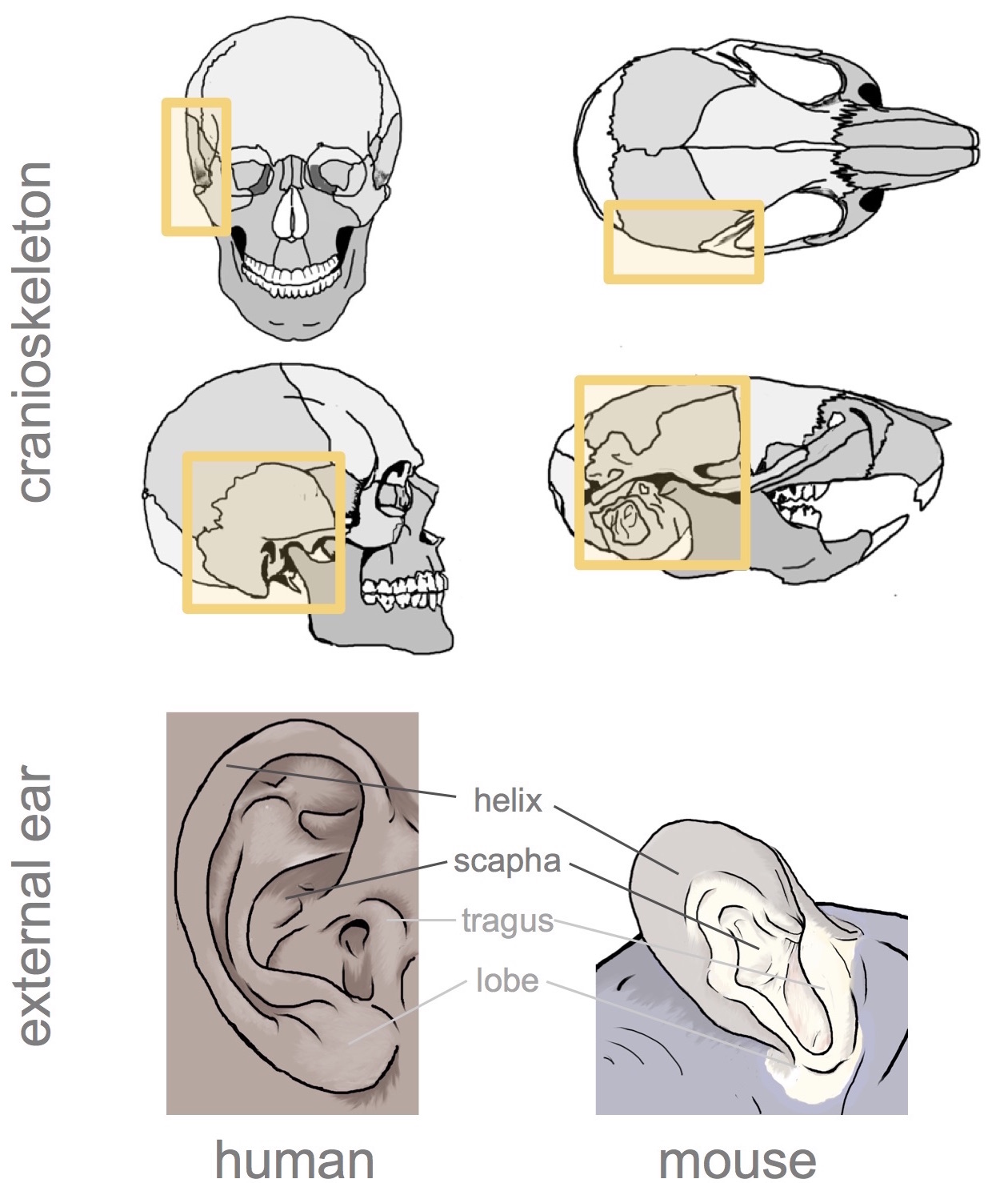
Originally published in Cox, et al. 2014
Cox TC, Camci ED,Vora SR, Luquetti DV, and Turner EE. The genetics of auricular development and malformation: New findings in model systems driving future directions for microtia research. Eur J Med Genet 2014;57.
Ossicles
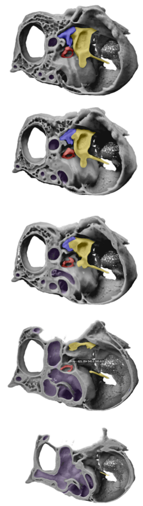
Pseudosections through a disarticulated tympanic bulla, showing the position of the ossicles, including the footplate of stapes.
Key
Yellow, incus
Blue, malleus
Red, stapes
Purple, inner ear</div>
Innervation
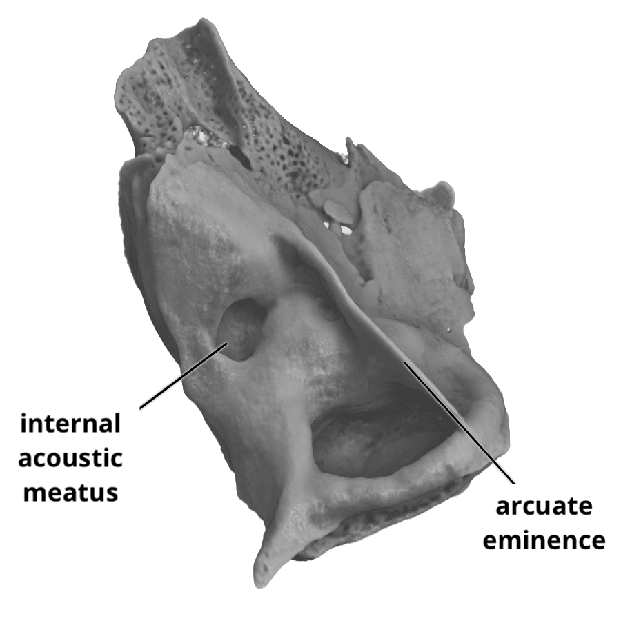
Rendering showing the endocranial surface of the tympanic bulla, with labels indicating the location of the arcuate eminence and the opening for the internal acoustic meatus.
The arcuate eminence is directly over the superior semicircular canal.
The cranial nerves VIII (vestibulocochlear) and VII (facial), as well as the labyrinthine branch of the cerebellar artery are transmitted through the internal acoustic meatus.